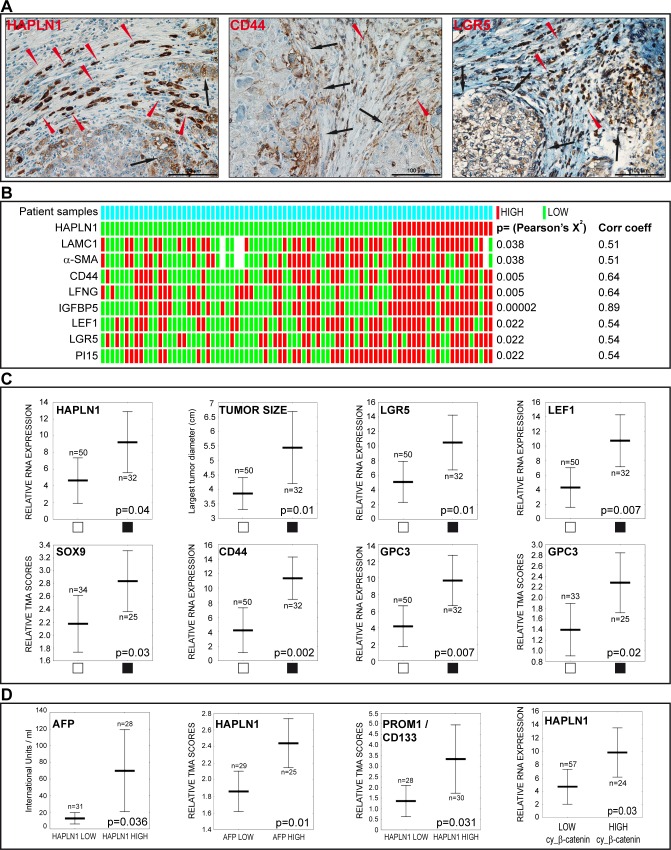
Figure 8. HAPLN1 is detected in HCCs featuring stem cell markers, high alpha-fetoprotein levels, vascular invasion and β-catenin activation.
A. Immunodetection of HAPLN1, CD44 and LGR5 in formalin-fixed, paraffin embedded HCCs. Brown color, specific signal; blue, hematoxylin counterstaining. The three proteins are detected in tumor cells at the tumor-stroma interface (black arrows) and in cells arranged in “Indian files” (red arrowheads). B. Heatmap showing associations of HAPLN1 mRNA expression with the indicated genes. White bars indicate missing data (n=8/729 spots). Pearson's X2 and Gamma correlation coefficients are shown on the right. C. Vascular tumor invasion (black squares) is associated with the indicated variables. D. Serum AFP levels are higher in HAPLN1 HIGH patients. High HAPLN1 protein correlates with high AFP serum levels. High PROM1/CD133 protein is detected in HAPLN1 HIGH HCCs. High HAPLN1 mRNA correlates with high cytoplasmic β-catenin.

