
Figure 1. Aging pathology in the C. elegans hermaphrodite gonad.
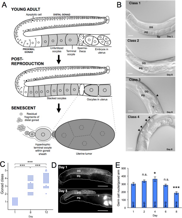
A. Schematic summary of major age changes in the anatomy of the hermaphrodite gonad, derived from previous sources [16, 18–20, 62, 63] and this study. Top, young, reproductive adult. Middle, post-reproductive adult, shortly after sperm depletion; note stacking of oocytes in proximal gonad and presence of oocytes in the uterus. Bottom, terminal state after pathogenetic changes in the aging gonad. Fragments comprised of clumps of cellularized germ cells are all that remains of the distal gonad, the terminal oocyte has undergone hypertrophy, and oocytes within the uterus have developed into a large tumor. B. Stages 1 - 4 of gonad deterioration; images from Nomarski microscopy. Class 3 arrowhead, narrowing of distal gonad. Class 4 arrowheads, distal gonad fragments. Scale bar, 20 μm. C. Age increase in gonad degeneration (wild type hermaphrodite). Each dot represents an individual animal. *** p < 0.001, Wilcoxon-Mann Whitney test. D., E. Numbers of germline nuclei decline with age. D. Gonads of young and old hermaphrodites stained with DNA-staining fluorescent dye (DAPI); scale bar, 20 μm. Note smaller number of nuclei in 8 day old worm. T, DNA mass in uterine tumor. PG, proximal gonad, DG, distal gonad. E. Age change in number of germline nuclei (stage 4, 5 worms on day 8 not included); * 0.01 < p < 0.05, *** p < 0.001, Student's t test, compared to day 1; error bars, S.E.M.; n.s., not statistically significant. N, sample size.