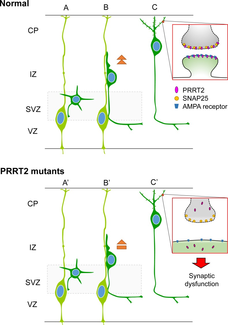
Figure 8. Schematic diagram showing effects of PRRT2 mutation on neuronal migration and dendritic spines in cortical neurons.
A. Radial glia cells (light green) produce postmitotic neurons (dark green). B. Postmitotic neurons then migrate along the radial fiber to the CP. C. After reaching the cortical plate, the cells extend dendrites and form synaptic connections with other neurons through dendritic spines. Box shows a magnification of the pre- and post-synaptic regions. PRRT2 (pink) is localized at both the pre- and post-synaptic membranes through interactions with SNAP2518, 25, 44 and the AMPA receptor25, 33, 34. (A', B') In neurons carrying PKD-associated PRRT2 mutations, reduced expression, mislocalization and other potential defects delay the neuronal migration. (C') PRRT2 mutation also decreases the dendritic spine density and causes synaptic dysfunction, possibly through SNAP25 and/or AMPA receptor pathways.

