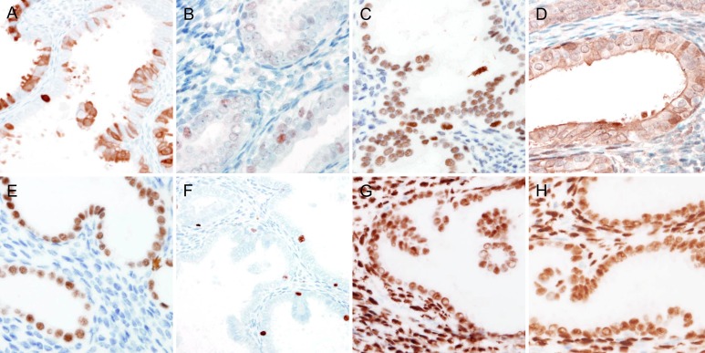
Figure 5. Immunohistochemical findings of endometrial papillary proliferation.
A. Patchy p16 immunoreactivity in the cytoplasm excluded the possibility of serous carcinoma. B. Faint to weak, focal p53 expression in a few epithelial cells indicated wile-type TP53, excluding the possibility of serous carcinoma. C. to E. In the epithelial cells of the endometrial papillary proliferation, the expressions of C. ARID1A, D. PTEN, and E. PAX2 were well preserved. F. Ki-67 labelling index was not significantly increased (less than 1%). G. and H. The epithelial cells of the endometrial papillary proliferation exhibited diffuse, strong immunoreactivity for G. estrogen receptor and H. progesterone receptor.

