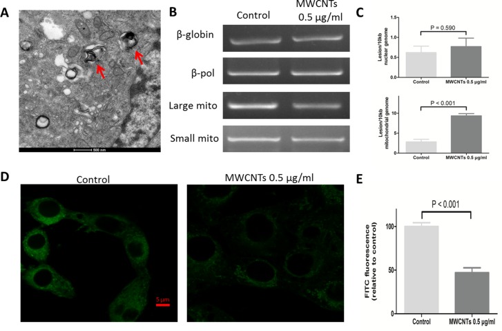
Figure 3. The effect of MWCNTs on the mitochondrial organelle in GC-2spd cells.
A. TEM image showed a section of GC-2spd cells treated with MWCNTs. MWCNTs were accumulated in mitochondria of GC-2spd. B. The electrophoretic results of nuclear genomic DNA fragments (i.e. nDNA) and mitochondrial genomic DNA fragments (i.e. mtDNA) in GC-2spd cells. C. Quantitative levels of damage to nDNA and mtDNA of the GC-2spd cells between MWCNTs treatment and controls were shown on each right side. D. The labeling of mitochondria in GC-2spd cells. Confocal microscopy images showing the mitochondria amount and location after MWCNTs exposure. E. Quantitative levels of mitochondria fluorescence intensity. Each data was represented as the means ± S.E. from three separate experiments.

