
Figure 1. 2D gel electrophoresis and western blot analysis of M. bovis HB0801 whole cell proteins.
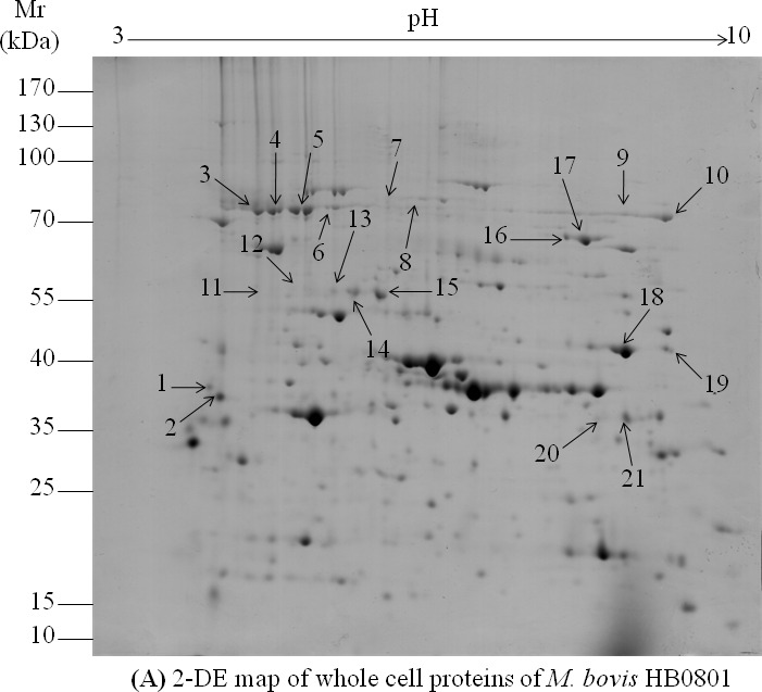
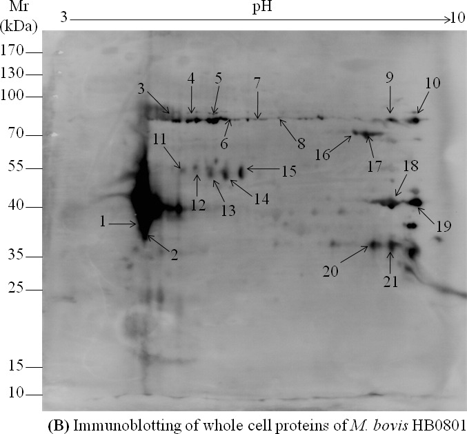
A. Analysis of HB0801 whole cell proteins with 2-DE. Isoelectric points are indicated at the top and molecular weight markers in kDa on the left. B. Immunoblotting patterns of M. bovis HB0801 whole cell proteins obtained using a pool of sera derived from calves experimentally infected with HB0801. The 21 spots identified by MALDI-TOF MS are indicated on the 2D electrophoresis gel and the PVDF membrane.