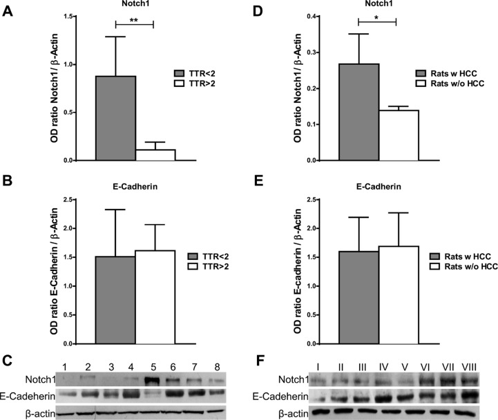
Figure 5. Notch1 and E-Cadherin expression in non-tumor liver tissue.
(A–B) Notch1 and E-Cadherin expression were evaluated by western blot in cirrhotic tissues surrounding HCC and a higher Notch1 expression was observed in cases with poor (< 2 years) vs. good (> 2 years) recurrence-free survival (TTR) **P < 0.01 (by two tailed student's t test). (C) Representative Notch1 and E-Cadherin expression in cirrhotic tissues with poor (< 2 years) (cases 5-8) vs. good (> 2 years) (cases 1-4) recurrence-free survival (D–E) Notch1 and E-Cadherin protein expressions were detected by western blot in liver of rats treated with DENA. Higher Notch1 expression was observed in non-tumor liver of rats that developed HCC (Rats w HCC) compared to those that did not developed (Rats w/o HCC). *P < 0.05 (by two tailed student's t test). (F) Representative Notch1 and E-Cadherin expressions in non-tumor liver of rats that developed HCC (cases V–VIII) compared to those that did not developed HCC (I–IV).

