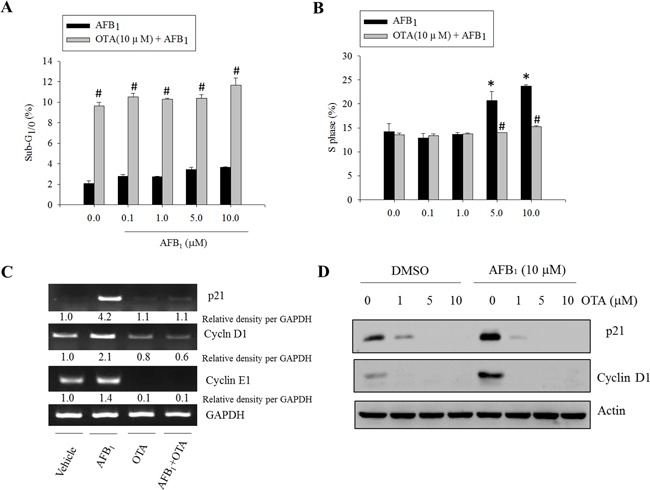
Figure 2. Effects of carcinogenic mycotoxins on the cell cycle in human intestinal epithelial cells.

HCT-8 cells were treated with different concentrations of AFB1 in the presence or absence of OTA (10 μM) for 24 h, and the cells were stained with PI for FACS analysis. A. The ratio of cells in the sub-G1/0 phase. B. The ratio of cells in the S phase. An asterisk (*) indicates a significant difference compared to the control group treated with DMSO alone (p < 0.05). A hatch mark (#) indicates a significant difference compared to the group treated with AFB1 alone (p < 0.05). C. HCT-8 cells were treated with AFB1 (10 μM), OTA (10 μM), or a combination of the two reagents for 24 h. mRNA expression of each gene was measured using real-time PCR. D. HCT-8 cells were treated with various concentrations of OTA in the presence or absence of AFB1. Total cell lysates were subjected to Western blot analysis.
