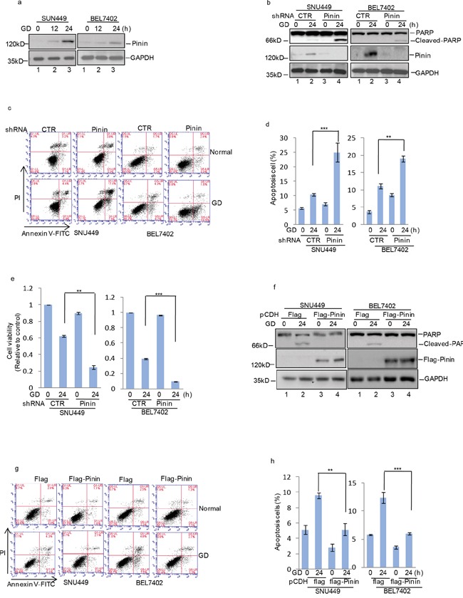
Figure 4. Pinin inhibits glucose deprivation–induced apoptosis in HCC cells.

a. SNU449 and BEL7402 cells were cultured with or without glucose deprivation for the indicated periods. Cell lysates were separated by SDS-PAGE and analyzed by western blot with indicated antibodies. GAPDH was used as the loading control. b. SNU449 and BEL7402 cells with or without knockdown of Pinin were cultured in present of glucose or not for 24 h. GAPDH was used as the loading control. c, d. The percentage of cell apoptosis was analyzed by flow cytometry analysis. (n=3, mean ± SD, t-test, **P<0.01, ***P<0.001 vs. shRNA CTR). e. Cell viability was determined by CCK8 assay.(n=3, mean ± SD, t-test, **P<0.01, ***P<0.001 vs. shRNA CTR). f. SNU449 and BEL7402 cells with or without overexpression of Pinin were cultured in present of glucose or not for 24 h. Cell lysates were then subjected to western blot analysis with the antibodies indicated. GAPDH was detected as the loading control. g, h. The percentage of cell apoptosis was analyzed by flow cytometry analysis. (n=3, mean ± SD, t-test, **P<0.01, ***P<0.001 vs. pCDH-flag).
