
Figure 4. Overexpression of BKCa stimulates prostate cancer cell proliferation, migration and invasion.
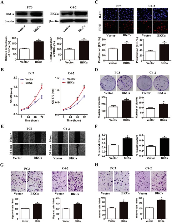
A. Prostate cancer PC3 and C4-2 cells were transfected with BKCa or Vector plasmids for 48 hr. The expression of BKCa was analyzed by western blot. β-actin was used as loading control. B. Cell growth curves as determined by MTT assay in PC3 and C4-2 cells transfected with BKCa or Vector. C. Percentage of EdU positive cells determined by EdU incorporation assay (lower panel) and representative images of EdU staining (upper panel) in PC3 and C4-2 cells transfected with BKCa or Vector. D. Effects of upregulated BKCa on the colony-genic ability of PC3 and C4-2 cells. Representative images were shown in the upper panel and the number of colonies was illustrated in the lower panel. E. Overexpression of BKCa promoted prostate cancer cell migration as determined by scratch wound healing assay. The representative images of wound healing were shown in left panel. The relative wound closure was illustrated in right panel. F-H. Effects of upregulated BKCa on the migration and invasion of PC3 and C4-2 cells as determined by transwell assays. Representative images and mean numbers of migrated (G) and invaded (H) cells were shown in the upper and lower panel respectively. All the experiments were performed in triplicate. The data are shown as the means ± se. *P < 0.05.