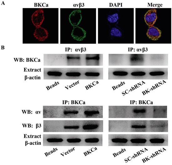
Figure 8. Characterization of the integrin αvβ3/BKCa complex in prostate cancer PC3 cells.

A. Immunocolocalization of BKCa and αvβ3. Representative confocal images of BKCa (red) and integrin αvβ3 (green) staining conducted in PC3 cells. Staining is more pronounced on the plasma membrane for both proteins and the merge shows that there is a co-localization (yellow to orange areas) on plasma membrane areas. B. Co-immunoprecipitation of integrin αvβ3 and BKCa in PC3 cells. Cells were transfected with BKCa or BK-shRNA and their corresponding negative control vectors. Proteins were extracted and immunoprecipitated using anti-BKCa or anti-αvβ3 antibodies; blots were probed with anti-αvβ3 or anti-BKCa antibodies respectively. Bead lanes contain the protein G conjugated sepharose beads used during the immunoprecipitation without the protein input. Equal amount of protein extract from each group was subjected to Western blot with β-actin as the loading control. All the experiments were performed in triplicate.
