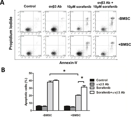
Figure 2. Blocking integrin αvβ3 increased leukemia sensitivity to sorafenib in BMSCs.

A. The apoptosis rate measured by flow cytometry by Annexin-V/PI double staining. MV4-11 cells were seeded onto the BMSC monolayer or cultured alone in 6-well plates with or without αvβ3 blocking antibody (1μg/ml) for 2 hours, sorafenib (10μM) was then added and cultured for 24 hours. Suspension cells were used for apoptosis assay. B. The percentage of apoptosis in each group. *p<0.05.
