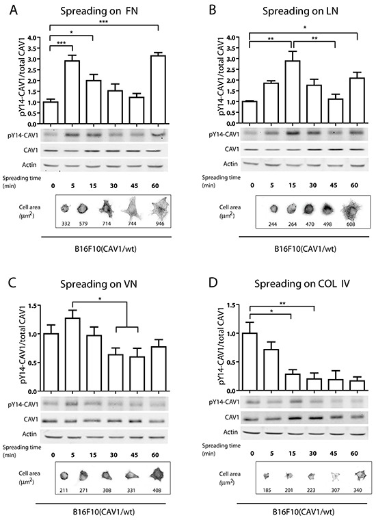
Figure 3. CAV1-phosphorylation on tyrosine 14 during cell spreading on pure ECM surfaces.

In spreading assays, B16F10(CAV1/wt) cells (1,5×10⁶) were allowed to attach to A. fibronectin, B. laminin, C. vitronectin and D. collagen IV-coated plates (2 μg/ml) for different periods of time (0, 5, 15, 30 and 60 min), with time 0 representing cells in suspension. Then, whole cell lysates were prepared and pY14-CAV1 levels were determined by Western blotting. Upper graphs show the densitometric analysis of relative pY14-CAV1 levels during cell spreading. Lower images show pY14-CAV1, CAV1 and Actin (control) expression by Western Blotting. Lower panels show cells in phase contrast and stained with phalloidin during spreading. The average area per cell is indicated in μm2. Data shown are the averages from three independent experiments (mean ± S.E.M, n=3,***p<0.001; **p<0.01 and *p<0.05).
