
Figure 1. miR-558 facilitates the translation of HIF-2α in NB cells.
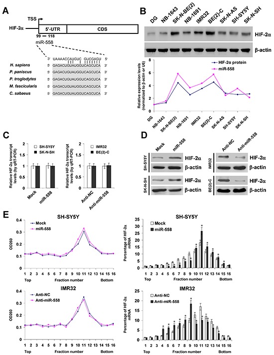
A. scheme of the potential binding site of miR-558 in the HIF-2α 5′-UTR, locating at bases 99-118 downstream the transcription start site (TSS). B. western blot and real-time qRT-PCR assays revealing the expression levels of HIF-2α and miR-558 in NB cell lines with [NB-1643, SK-N-BE(2), NB-1691, IMR32, BE(2)-C] or without MYCN amplification (SK-N-AS, SH-SY5Y, SK-N-SH) and normal dorsal ganglia (DG). C. and D. real-time qRT-PCR and western blot assays showing the transcript and protein levels of HIF-2α in NB cells transfected with empty vector (mock), miR-558 precursor, negative control inhibitor (anti-NC, 100 nmol/L), or anti-miR-558 inhibitor (100 nmol/L). E. sucrose gradient sedimentation assay indicating the distribution of HIF-2α transcripts to polysome fractions in NB cells transfected with mock, miR-558 precursor, anti-NC (100 nmol/L), or anti-miR-558 inhibitor (100 nmol/L). * P<0.01 vs. mock or anti-NC.