
Figure 7. High expression of HIF-2α is positively correlated with miR-558, AGO2, or eIF4E levels in NB tissues.
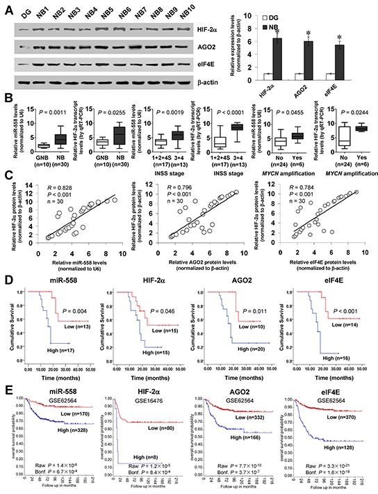
A. western blot indicating the expression of HIF-2α, AGO2, and eIF4E in NB tissues (n=30) and normal dorsal ganglia (DG). B. real-time qRT-PCR assay showing the expression of miR-558 and HIF-2α in ganglioneuroblastoma (GNB, n=10) and NB (n=30), and their levels in NB cases with different INSS stage or MYCN amplification status. C. the correlation between HIF-2α protein and miR-558 transcript, AGO2 protein, or eIF4E protein levels in NB tissues (n=30). D. and E. Kaplan–Meier survival plots of 30 well-defined NB cases (stratified by median value) and public NB cohorts (stratified by the scan method and adjusted by Bonferroni correction) derived from GEO database and R2 microarray analysis and visualization platform (http://r2.amc.nl) indicating the survival probability of patients with high or low expression of miR-558, HIF-2α, AGO2, or eIF4E. * P<0.01 vs. DG.