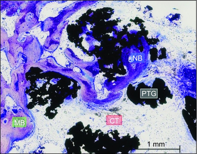
Figure 7.
Close view Group A. Connective tissue was present between particles, with some bone formation around and inside some granules; however, other particles were surrounded only by connective tissue. The particles were mainly visible in the vicinity of the defect walls, not in the medullar area, most likely as result of particle movement and primary connective tissue formation. Toluidine blue staining, magnification ×15. CT, connective tissue or its space; MB, mature bone; NB, new bone; PTG, porous titanium granule.

