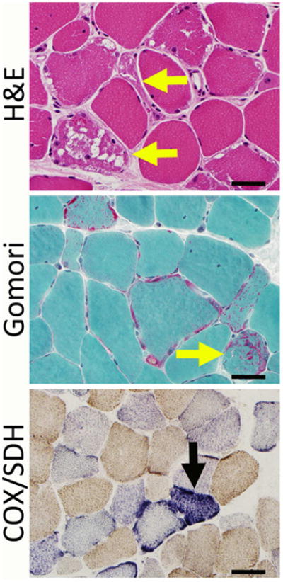
Figure 3. Pathological Findings in Severe MD.

Severe mitochondrial disease can show alterations in fiber size, ragged red fibers (yellow arrows), and accumulations of mitochondria (here seen as red-stained subsarcolemmal areas on the Gomori trichrome stain). COX and SDH stains may show stains that are negative for COX, and some of these fibers may overexpress SDH (black arrow). Bar = 40 mm.
