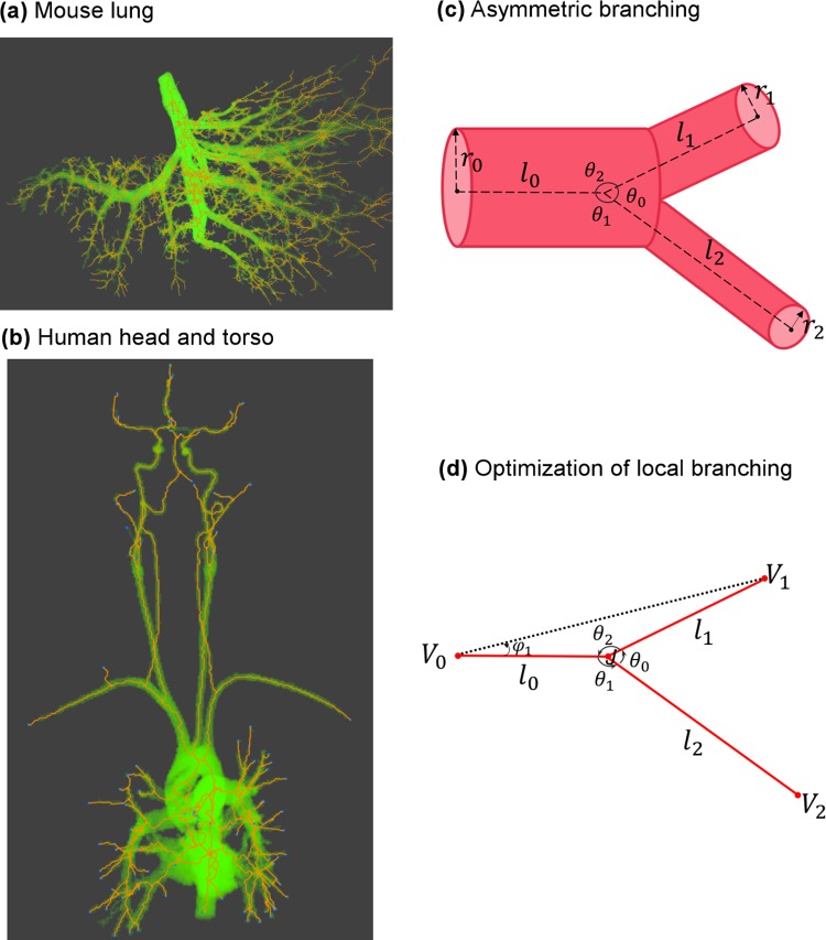
Fig 1. Cardiovascular data and schematic illustration of vascular branching.
(a) Mouse lung micro-CT images processed by Angicart. (b) Human head and torso MRI images processed by Angicart. (c) Schematic illustration of the asymmetric branching geometry and labeling. Parent vessel with radius r0 and length l0 branches into two daughter vessels with radius ri and length li with subscript i = 1 or 2. Branching angles, θi, are defined by the angle between the sides defined by the endpoints of the vessel pairs. Here, subscripts are determined by the non-adjacent vessel. (see Materials and Methods) (d) Optimization of local branching on a plane finds the optimal location of the branching junction J when the unshared endpoints (Vi) and the radii (ri) are fixed (see General framework for branching angle optimization and asymmetry).

