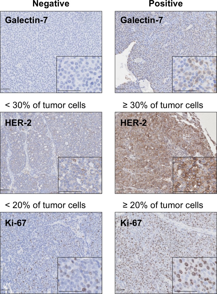
Fig 3. Immunohistological analysis of mammary tumour markers in presence or absence of gal-7.
Immunohistochemical staining showing membrane-bound HER-2-positive cells and nuclear Ki67 expression in mammary tumors that were negative (left panels) or positive (right panels). Scale bars, 50 μm and 25 μm (insets).

