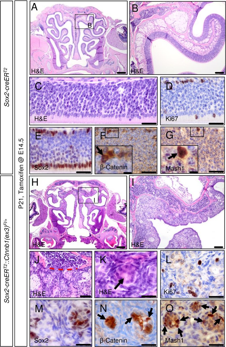
Fig 3. Embryonically activated aberrant Wnt signaling in Sox2-creERT2::Ctnnb1(ex3)Fl/+ mice leads to the formation of tumor-like lesions within the olfactory epithelium.
Tamoxifen application in Sox2-creERT2::Ctnnb1(ex3)Fl/+ mice at E14.5 causes aberrant activation of the canonical Wnt signaling pathway in Sox2-positive cells of the mouse OE and leads to the formation of tumor-like lesions within this structure. As shown for P21 mice by H&E staining, the structure of the native OE in mutant mice compared to controls is fully disrupted. Alterations are more pronounced in the upper nasal cavity (A, OE of control mice; B, higher magnification of framed area in A; H, OE tumor-like lesions of mutant mice; I, higher magnification of framed area in C). OE tumor-like lesions of mutant mice display signs of infiltration with disruption of bone laminae (C, control OE; J, red dashed line indicates broken bone barrier in mutant OE lesions) as a feature of malignant growth. Rosette-like structures are present in OE lesions of mutants (C, control OE; K, rosette in mutant OE lesion indicated by arrow). The amount of Ki67-positive cells in mouse OE tumor lesions remains comparable to the native OE (D, control OE; L, mutant OE lesion). Staining for Sox2, ß-Catenin and Mash1 delineate tumor cell nests from a surrounding stromal cell compartment (E, F, G, control stains; M, N, O, stains in mutant OE lesions). Scale bars equate to 500 μm in A and H, equate to 100 μm in B and I, equate to 25 μm in C-G and J-O and equate to 5 μm in insets.

