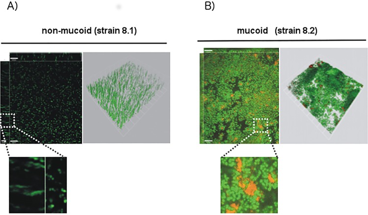
Fig 4. Confocal microscopy of biofilms stained with PIA/PNAG-specific antibodies.
Non-mucoid strain (A) and mucoid (B) S. aureus strains (strains 8.1 and 8.2) were grown overnight in TSB. After washing, adherent bacteria were stained using SYTO 9 (green). PIA/PNAG was detected using ALEXA-568 labeled wheat germ agglutinin (red), which is a lectin binding to the N-acetylglucosaminyl backbone of PIA/PNAG. Zoom-in shows PIA/PNAG-embedded bacteria in the mucoid strain (B), while hardly any PIA/PNAG is detected with the non-mucoid isolate. White bar = 10 μm.

