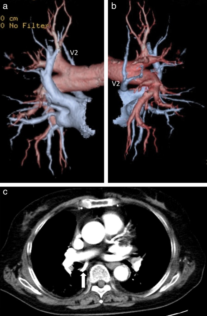
Figure 3.

Abnormal distribution of the aberrant pulmonary vein (V2) descended dorsally and emptied into the left atrium. (a) Front view; (b) right side view. (c) V2 (white allow) behind the right main bronchus independently drained directly into the left atrium, observed on two‐dimensional computed tomography.
