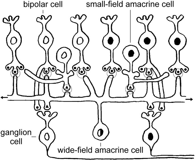
Fig. 10.
Schematic circuit for generating mean rate changes in ganglion cells during remote stimulation. The circuit assumes that amacrine cells form inhibitory chemical synapses (Pourcho and Goebel, 1983). As such, wide-field ON–OFF amacrine cells (black-and-white nucleus) rectify input from ON- and OFF-center (white and black nuclei, respectively) bipolar cells and small-field amacrine cells within their dendritic tree and transmit the pooled result to ON- and OFF-center ganglion cells throughout the retina via their long axon-like processes. The transmission is partially mediated by spikes (Cook et al., 1998; Demb et al., 1999); electrical connections between wide-field amacrine cells may also be involved (Naka and Christensen, 1981; Kolb and Nelson, 1985). This wide-field amacrine cell pathway would constitute the nonclassical receptive field of ganglion cells. The classical receptive field presumably derives from the bipolar–ganglion cell pathway depicted in the figure. Filled circles and triangles indicate excitatory and inhibitory synaptic transmission, respectively. Dotted circles indicate rectifying synapses. Note that the somata of wide-field amacrine cells are found in the inner plexiform and inner nuclear layers in addition to the ganglion cell layer (Stafford and Dacey, 1997).

