
Fig. 8.
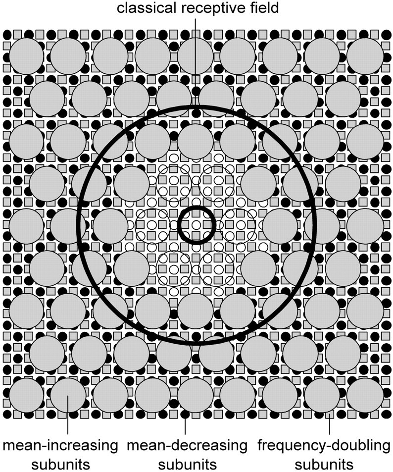
The ganglion cell receptive field. This field consists of the familiar center-surround mechanisms (thick lines) and three nonlinear mechanisms. These mechanisms are more potent in Y-cells than in X-cells. They appear to operate by rectifying input from large or small subunits distributed over widespread regions of the retina. For purpose of illustration the figure depicts individual subunits, but they should be collectively viewed as a sheet because no one of them can evoke a response from the cell. The large subunits subserve a mechanism that acts to increase the mean rate during continuous background stimulation (large gray circles), whereas, the small subunits appear to subserve two mechanisms, one that acts to decrease mean rate during such stimulation (small black circles) and one known from previous work to generate frequency-doubled responses to back-and-forth movements of large patterns (small gray squares). The latter mechanism may also contribute to mean rate changes, but its contribution would be small compared with the mean-decreasing mechanism, at least for stimuli moving outside the classical receptive field. The various subunits are shaded gray orblack to indicate whether they have an excitatory or inhibitory effect on ganglion cells, respectively. Those not shaded are presumed to exist, but we could not probe the central portion of the receptive field to confirm this (see Results for explanation). All receptive field elements are drawn to scale for a Y-cell. For reference, the small subunits are 0.3° in diameter. Their surrounds are not depicted in the figure.