Fig. 3.
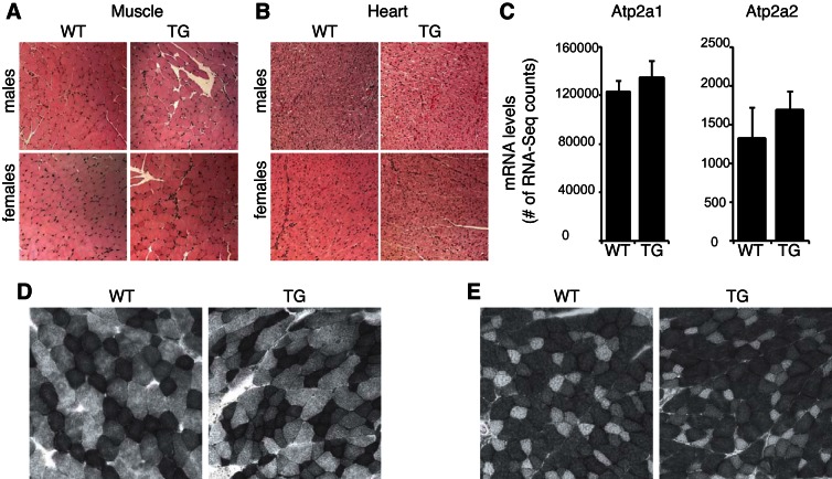
No morphological differences in muscle and heart tissue between ZFP-TG and WT mice. A and B: muscle (A) and heart tissue (B) from ZFP-TG (TG) and WT mice stained with hemotoxylin and eosin (n = 4 mice/group). Total magnification, ×200. C: gene expression of Atp2a1 (fast-twitch muscle fiber marker) and Atp2a2 (slow-twitch muscle fiber marker) in muscle from TG and WT male mice (n = 5/group). D and E: representative succinate dehydrogenase (D) and α-glycerol-3-phosphate dehydrogenase staining (E) from muscle of ZFP-TG and WT male mice (n = 3/group).

