Fig. 1.
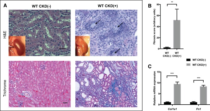
Tubulointerstitial inflammation, crystalline deposits, and interstitial fibrosis in the kidneys of developing adenine-fed mice. A: macroscopic appearance (insets) and histology of the kidneys. Control kidney appears normal, whereas kidney of an adenine-fed mouse is pale, with a rough tuberous surface. Hematoxilin and eosin (H&E) staining (top, ×40 magnification) demonstrated crystalline deposits (arrows) and severe tubulointerstitial inflammation in the kidneys of WT (wild-type) mice with chronic kidney disease [CKD(+)]. Mason trichrome staining (botttom, ×20 magnification) revealed advanced interstitial fibrosis, with tubular loss and atrophy in the kidneys of adenine-fed CKD(+) mice. Scale bars: 20 μm, top; 50 μm, bottom. B: fibrosis quantification with image deconvolution technique. Adenine fed mice had 40-fold more Mason trichrome blue stained collagen compared with control mice (4 mice per group). C: collagen type I-α1 (Col1a1) relative mRNA expression was 9-fold higher, and fibronectin 1 (Fn1) mRNA 8-fold higher in the kidneys of adenine-fed mice compared with controls. Three kidney samples per group were used for PCR. Error bars represent SD. **P < 0.01; ***P < 0.001.

