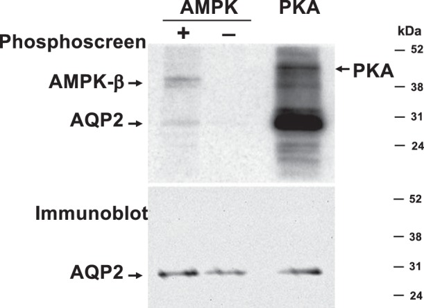
Fig. 4.

AMPK fails to significantly phosphorylate AQP2 in vitro. V5-tagged AQP2 was expressed in HEK-293 cells, immunoprecipitated, and incubated with [γ-32P]ATP in the presence of PKA catalytic subunit (positive control) or in the presence or absence of AMPK holoenzyme. A phosphoscreen image (top) and immunoblot (bottom) of the same membrane are shown (representative of 4 experiments). AQP2 gets robustly phosphorylated in the presence of PKA, but only weakly phosphorylated in the presence of AMPK (top). PKA catalytic subunit and the AMPK β-subunit get autophosphorylated, as indicated. The immunoblot (bottom) reveals similar AQP2 loading in all lanes.
