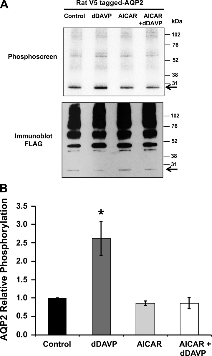
Fig. 6.
AMPK activator blocks vasopressin-induced phosphorylation of AQP2 in mpkCCDc14 cells. mpkCCDc14 cells were transiently transfected with V5-tagged rat wild-type AQP2 plasmid. Twenty-four hours posttransfection, cells were incubated in the presence or absence of AICAR (2 mM) for 20 h, and then dDAVP (10 nM) or vehicle was added during the last 30 min of the incubation. [32P]orthophosphate labeling of cells before cell lysis, immunoprecipitation, SDS-PAGE, and immunoblotting and phosphoscreen exposure of the same membrane were then performed as described in materials and methods. A: typical phosphoscreen image (top) revealing the signal of AMPK in vivo phosphorylated AQP2. The immunoblots (bottom) confirm similar protein expression and loading of the gel for the different conditions. B: quantification of AQP2 phosphorylation signal normalized for protein loading, as assessed by densitometry of the immunoblot. While AMPK activation alone did not induce significant changes in AQP2 phosphorylation, treatment with the AMPK activator AICAR prevented the dDAVP-induced increase in AQP2 phosphorylation. Values are means ± SE of 4 independent experiments. *P < 0.05 relative to the vehicle control.

