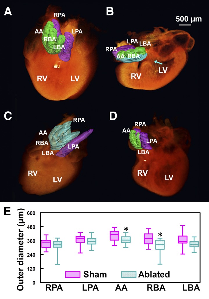
Fig. 1.
Cardiac neural crest cell (CNCC)-related great vessel defects. A–D: 3-dimensional (3D) optical coherence tomography (OCT) volume reconstructions and great vessel segmentation. Green, aortic trunk vessels; purple, pulmonary trunk vessels; cyan, branching errors. A: control embryonic heart. B: an example of persistent truncus arteriosus (PTA). Cyan-colored region at the great vessels represents common trunk; arrow points to ventricular septal defect. C: example of incorrect branching. Aortic trunk branches into 4, instead of 3, vessels, and pulmonary trunk branches into 1, instead of 2, vessels. D: absent great vessel. E: box plot of great vessel outer diameter measurements. Box represents 25–75%; whiskers represent range. Aortic arch (AA) and right brachiocephalic artery (RBA) are significantly smaller in CNCC-ablated than control group. *P < 0.05. LBA, left brachiocephalic artery; RPA, right pulmonary artery; LPA, left pulmonary artery; LV, left ventricle; RV, right ventricle.

