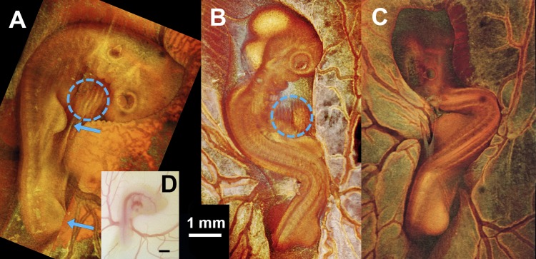
Fig. 3.
Whole embryo images demonstrating body flexure [Hamburger and Hamilton (HH) stage 19–20]. A–C: projection images of a sham control embryo (A) and 2 CNCC-ablated embryos (B and C) from 3D OCT volume rendering. D: microscope photo of embryo shown in A. Blue arrows point to limb buds that were used for staging embryos. Blue dashed-line circles, embryonic hearts (with artifacts due to the beating motion). Heart in C is not visible. Limb morphology indicates that all 3 embryos are at HH stage 19–20, closer to HH stage 20.

