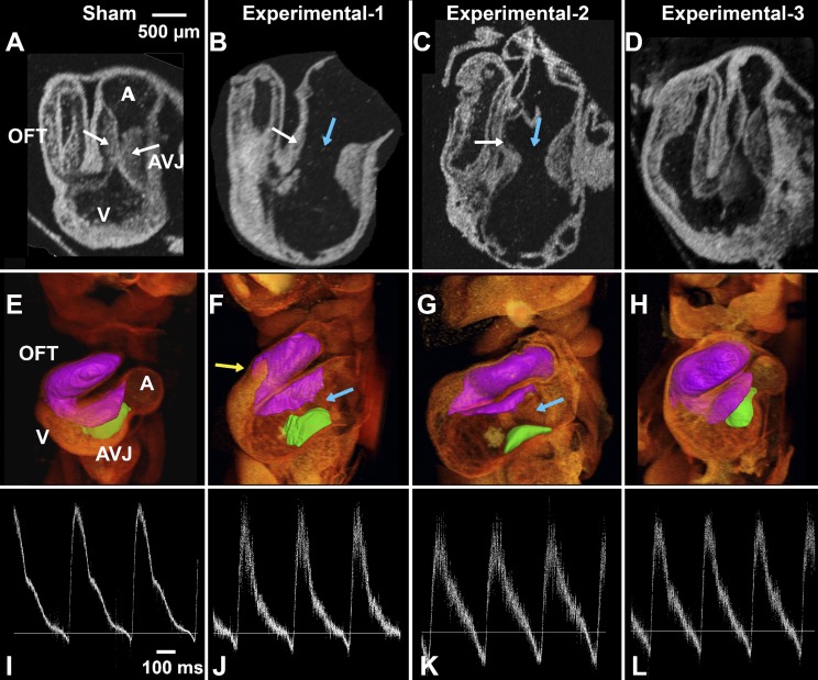
Fig. 5.
CNCC-ablated embryos have abnormal endocardial cushions at HH stage 19–20. A–D: 2-dimensional OCT sections of HH stage 19–20 embryonic hearts. E–H: 3D OCT volume rendering and cushion segmentations [inferior AV cushion (green) and fusion of superior AV cushion and outflow tract cushions (purple)] of HH stage 19–20 embryonic hearts. I–L: flow traces recorded in vivo. Panels in each column show data from the same embryo in vivo (pulse Doppler trace) and ex vivo (3D heart structural imaging) as labeled on the top of each column. A, E, and I: sham control embryo; B–D, F–H, and J–L: 3 experimental embryos. White arrows, superior and inferior cushions (thick cushions that touch each other in the control embryo and thin cushions in ablated embryos); blue arrows, large gaps between superior and inferior AV cushions in the ablated embryos (B, C, F, and G); yellow arrow, reduced cushion coverage in an ablated embryo. D: CNCC-ablated embryo that appeared to have relatively normal cushions. A, atrium; AVJ, AV junction; V, ventricle; OFT, outflow tract.

