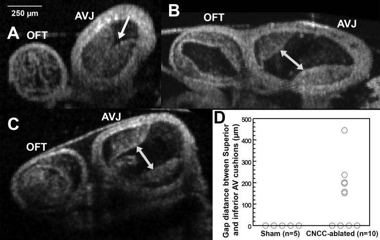
Fig. 6.
OCT cross-sectional images of AV cushions. A: control embryonic heart. B and C: CNCC-ablated embryonic hearts. Arrow in A, inferior and superior AV cushions contact each other and fill most of the lumen; double-headed arrows in B and C, gaps between inferior and superior AV cushions in ablated hearts. D: gap distance measurements. All 3 images share the same scale bar on the top left corner.

