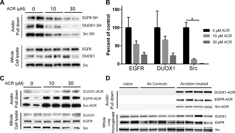
Fig. 7.
ACR exposure induces cysteine modification and carbonylation within DUOX1, EGFR, and Src. A and B: untreated or ACR-treated HBE1 cells (30 min) were lysed in the presence of the thiol-specific reagent EZ-Link Iodoacetyl-LC-Biotin, and avidin-purified proteins as well as whole cell lysates were analyzed by Western blot for DUOX1, EGFR, and Src. A: representative blots. B: densitometry analysis of relative thiol content of DUOX1, EGFR, or Src from 2 experiments in duplicate. *P < 0.05. C: untreated or ACR-treated HBE1 cells (30 min) were derivatized with ARP, and avidin-purified proteins or whole cell lysates (input controls) were evaluated by Western blot for DUOX1, EGFR, or Src. D: mice were exposed to ACR (5 ppm; 4 h) or filtered air, and lung homogenates were derivatized with biotin hydrazide for similar analysis of carbonylated proteins. Representative blots from 2–3 separate experiments are shown.

