Fig. 1.
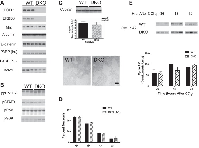
Creation of a hepatocyte-specific epidermal growth factor receptor-ERBB3 double knockout (HS-EGFR-ERBB3 DKO). A and B: EGFR and ERBB3 were genetically ablated in mice and evaluated by immunoblot for the expression of a number of proteins (A) or phosphorylated proteins (B). Of the proteins examined, the only proteins that changed in the livers of adult male mice were EGFR and ERBB3. PARP, poly(ADP-ribose) polymerase; in, intact; cl, cleaved. C: expression of cytochrome P450 2E1 (Cyp2E1) was analyzed in immunoblots of liver lysates from wild-type (WT) and DKO mice. As shown in the immunoblot and densitometric analysis, no difference was seen in the overall expression of this enzyme, which is required for CCl4-induced liver damage. The photomicrograph shows a representative section of the centrilobular damage caused by CCl4. The lighter areas around central veins show the loss of hepatocytes attributable to necrosis and apoptosis. Bar = 50 μm. D: mice were injected with CCl4 at 0 time and monitored for liver damage at 24, 48, 72, and 96 h after injection. No differences were observed in the necrotic area at 24 h, indicative that the initial necrotic areas were comparable in WT and DKO mice. The rate of “wound shrinkage or healing” was also comparable over the next 96 h. E: lysates from the livers of WT and DKO mice injected with CCl4 were monitored by immunoblots (left) for cyclin A2, a marker of DNA synthesis. This marker was not detected in control mice at zero time (data not shown). No differences were seen between WT and DKO mice in quantitative densitometry of cyclin A2 in DKO or WT mice (right).

