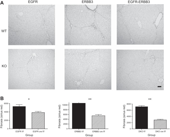
Fig. 2.
Analysis of fibrosis in WT and DKO mice subjected to repeated CCl4 injection over a 4-wk period. A: we injected HS-EGFR, HS-ERBB3, HS-EGFR-ERBB3 knockout mice, and their respective WT controls with CCl4 biweekly over 4-wk period. This resulted in fibrosis, as shown by staining liver sections with Sirius red to detect fibrillar collagen. Olive oil-injected controls showed staining mainly in the portal triads (data not shown). In contrast, both WT and KO livers showed fibrous strands, which sometimes bridged between central veins. These strands were decreased in all ERBB KO groups, particularly in the DKO mice. Bar = 100 μm. B: we quantified the fibrous area (excluding the normal areas of fibrosis around the portal triads) and confirmed the histologic impression of a significant reduction in fibrosis in the KO mice compared with their WT counterparts, particularly the DKO mice. All bars in graphs represent means + SE. *P < 0.01; **P < 0.0001.

