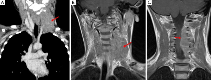
Figure 11.
This patient has a primitive neuroectodermal tumor which presented as a neck mass, this is demonstrated in image A (Coronal CT Neck) as a mass which is isodense to surrounding muscle (arrow) and on T1 weighted MRI (image B) this structure demonstrates an intermediate signal. Image C is also from a T1 weighted MRI acquisition and shows the tumor to be encroaching on the spinal canal and causing cord compression.

