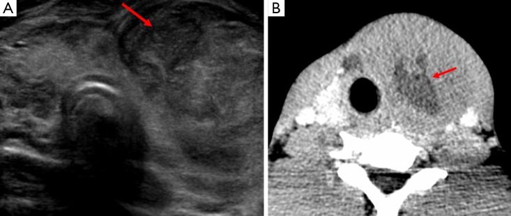
Figure 14.
Image A is a short axis view of the thyroid on ultrasound, it demonstrates a large complex mass within the left lobe of the thyroid (arrow) with a mixed pattern of internal echogenicity and some posterior acoustic enhancement. There is evidence of slight lateral deviation of the trachea which is confirmed on image B, which is the CT from the same patient. Image B also demonstrates areas of central hypoattenuation secondary to necrosis (arrow) and there is loss of surrounding fat planes. Although not demonstrated on this image there was also compression of the trachea and adjacent carotid artery and jugular vein. This was later diagnosed as non-Hodgkin’s lymphoma of the thyroid gland.

