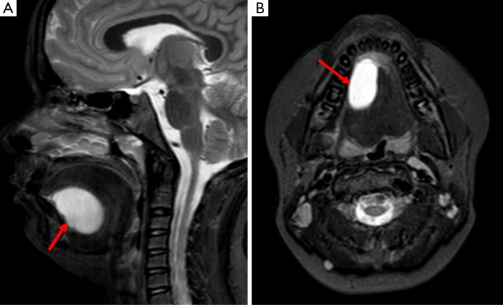
Figure 17.
Images A and B are sagittal and axial STIR sequences respectively of the floor of the mouth in a 10-year old child; they demonstrate a well-defined lesion of high (fluid) signal in the sublingual space (arrows). It was of low signal on T1 and demonstrated free diffusion on diffusion weighted imaging; overall this is in keeping with a ranula. If there was peripheral enhancement after gadolinium had been administered, this would raise the suspicion of infection.

