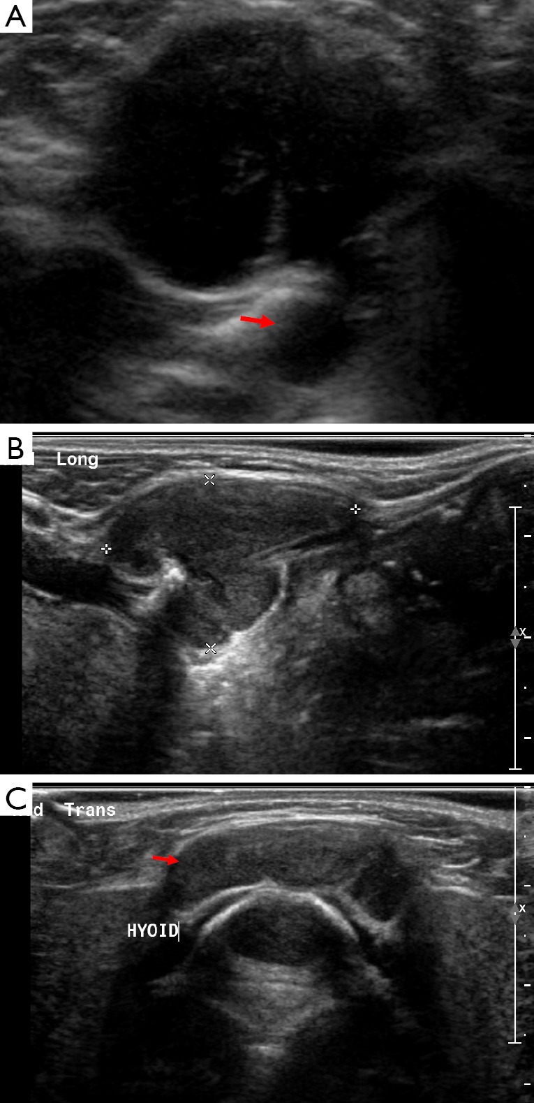
Figure 2.

Image A is an ultrasound showing a well-defined hypoechoic structure in the neck at the level of the hyoid, there is posterior acoustic enhancement and a tract connecting it with deeper structures (arrow). On Doppler imaging there was no increased internal vascularity. The imaging characteristics and midline location are consistent with the diagnosis of a thyroglossal duct cyst. Images B and C are of the same child who represented when the thyroglossal duct cyst became infected, note how the internal echogenicity has altered.
