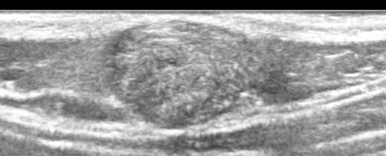
Figure 8.

This is an ultrasound image of a superficial mass in an 8-year old child on the back of his neck which demonstrates mixed internal echogenicity with a peripheral rim of low echogenicity; on doppler imaging this did not demonstrate increased internal vascularity; there are some internal hyperechoic foci that suggest possible calcification; overall the appearances are consistent with a pilomatrixoma.
