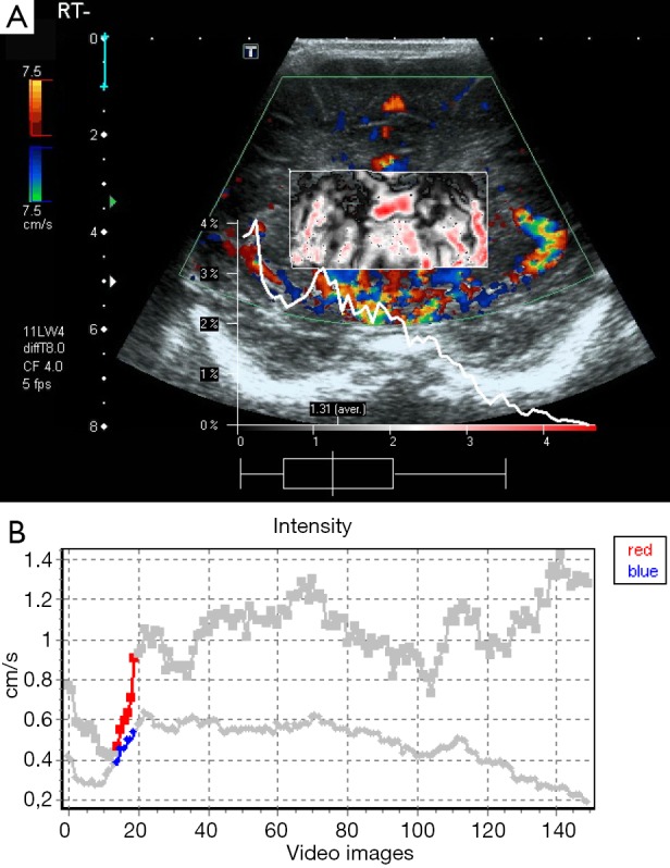
Figure 2.

A 1-day-old male infant with severe HIE. Dynamic tissue perfusion measurement demonstrates color Doppler image with ROI in the basal ganglia and very increased perfusion (A). Corresponding perfusion intensity curve with perfusion up to 0.9 cm/s (B). HIE, hypoxic-ischemic encephalopathy; ROI, region of interest.
