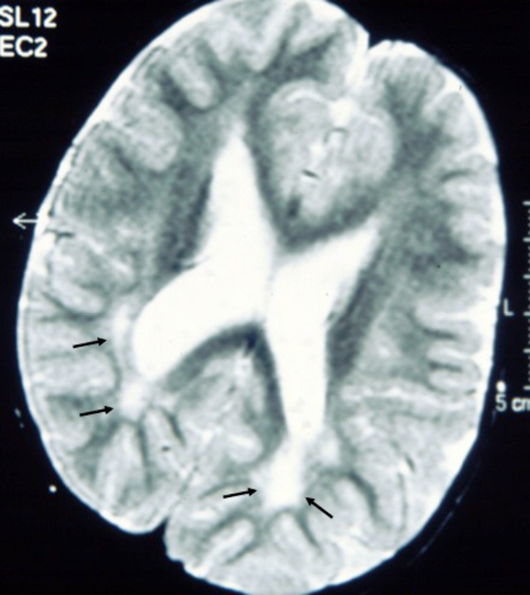
Figure 3.

Axial T2-weighted magnetic resonance images of the brain in a child with syndromic cutis tricolor showing high signal lesions in the posterior (peritrigonal) white matter (black arrows): thes hyperintensities were recorded at age 6 years and regarded as delayed myelination.
