Figure 2.

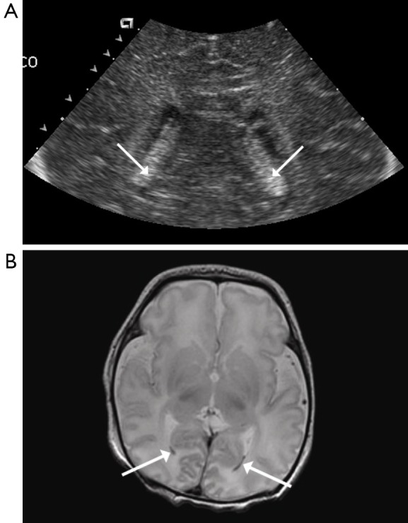
Bilateral IVH. (A) HUS with coronal image shows symmetrically enlarged choroid plexus (arrows); (B) MRI with T2 weighted axial image confirming bilateral IVH (arrows). HUS, head ultrasound; IVH, intraventricular hemorrhage.

Bilateral IVH. (A) HUS with coronal image shows symmetrically enlarged choroid plexus (arrows); (B) MRI with T2 weighted axial image confirming bilateral IVH (arrows). HUS, head ultrasound; IVH, intraventricular hemorrhage.