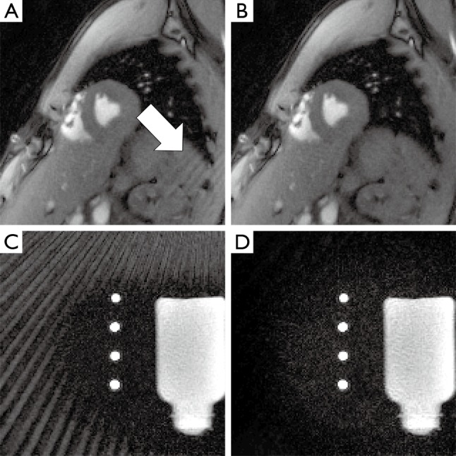
Figure 4.

Representative frames of MRI movies of (A,B) a cardiac short-axis view and (C,D) a phantom with static water and perpendicular flow in four tubes. (A,C) Reconstructions using all coils and (B,D) of a subset of coils after sinogram-based selection. In the cardiac study streak artifacts originate from the shoulder. For the flow phantom, a larger tube with back flow above the actual FOV generates strong streak artifacts. In both cases, the artifact intensity is greatly reduced by sinogram-based coil selection. MRI, magnetic resonance imaging; FOV, field of view.
