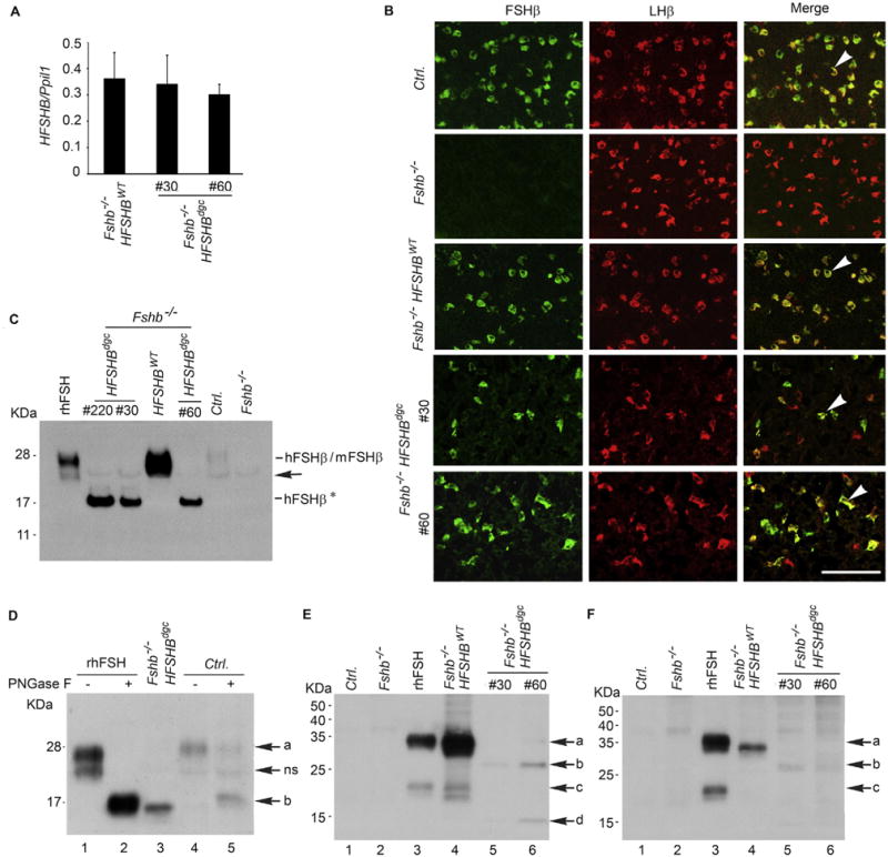
Fig. 2.

Real time qPCR assay (A) shows nearly identical expression of HFSHB mRNA in pituitaries of Fshb−/− mice expressing either a HFSHBWT or HFSHBdgc transgene. Two different lines #30 and #60, expressing the mutant transgene were developed. Dual label immunofluorescence visualization of pituitary sections (B) from adult mice confirmed gonadotrope-specific expression of HFSHBdgc transgene similar to that of HFSHBWT transgene (indicated by white arrowheads in merged images in the right panel in B). The absence of any green fluorescence in pituitary sections from Fshb−/− mice confirmed the specificity of the FSHβ antibody. The white bar in panel B represents 50 µm. Western blot analysis of proteins under denaturing conditions probed with a goat anti-human FSHβ-polyclonal antibody obtained from Santa Cruz Biotechnology, sc 7757 (C) shows a 15–17 KDa hFSHβAsn7Δ Asn24Δ non-glycosylated subunit (denoted as hFSHβ*) was abundantly expressed in pituitaries of three different lines of HFSHBdgc transgenic mice (line # 220, line #30 and line #60) on an Fshb null genetic background. Black arrow indicates a non-specific band. Recombinant human (r–h) FSHβ size-matched with that of HFSHBWT transgene-encoded hFSHβ subunit and weakly immunoreactive mouse (m) FSHβ subunit detected in normal control mouse (Ctrl.) pituitary. Proteins from Fshb−/− mouse pituitaries served as a negative control. PNGase F treatment followed by Western blot analysis of either r-hFSH or pituitary proteins from a normal control wild-type (Ctrl.) mouse showed an identical size de-glycosylated FSHβ subunit corresponding to that identified in an Fshb−/− HSFHBdgc mouse pituitary (D). Electrophoretic separation of proteins under non-denaturing conditions followed by Western blot analysis showed that in both male (E) and female (F) Fshb−/− HSFHBdgc mouse pituitaries, the non-glycosylated FSHβ subunit, despite high level expression (C) was present as an interspecies hybrid (mouse α + human FSHβ*) heterodimer (lanes 5 and 6 in panels E and F) at very low levels compared to the FSH heterodimer (mouse α + hFSHβ) in pituitaries of Fshb−/− HSFHBWT mice (lane 4 in panels E and F). Pituitary proteins from control and Fshb−/− mice (Lanes 1 and 2 in panels E and F) are used as negative controls and r-hFSH (Lane 3) as a positive control. Arrows in panel D denote: a = FSHβ subunit, ns = non-specific band, b = deglycosylated FSHβ subunit. Arrows in panel E and F indicate: a = FSHβ subunit, b = double N-glycosylation site mutant FSHβ subunit, c = uncombined/free FSHβ subunit, d = uncombined double N-glycosylation site mutant FSHβ subunit. (For interpretation of the references to color in this figure legend, the reader is referred to the web version of this article.)
