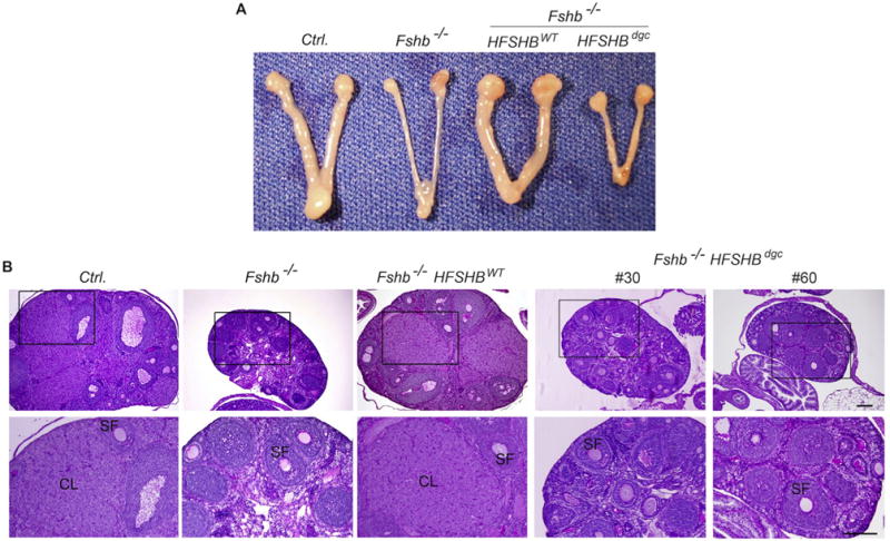
Fig. 5.

Gross morphology (A) and PAS-hematoxylin stained ovarian histology (B) showed that HFSHBdgc transgene unlike the HFSHBWT transgene did not rescue Fshb−/− females. Multiple developing secondary follicles and corpora lutea are readily seen in ovarian sections obtained from control (Ctrl.) and Fshb−/− HFSHBWT mice but not in those from Fshb−/− or Fshb−/− HSFHBdgc mice. Boxed areas in top panel in B are enlarged and shown below. SF: secondary follicle; CL: corpus luteum. The bar in panel B represents 200 μm.
