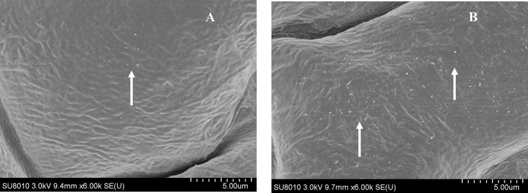
Figure 5.
Field Emission Scanning Electron Microscopy (FESEM) analysis of cuticle wax depositions. Adaxial side of 4-week-old Arabidopsis rosette leaves of WT and OE were observed under 6000 × magnification. (A) WT showed only little wax deposition on leaf surface. (B) The leaf surfaces of transgenic plants with high wax deposition. Wax crystals were shown by arrows.

