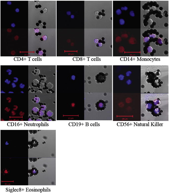
Fig. 3.

Nuclear morphologies of isolated cell types. Isolated leukocytes, bound to Dynabeads were stained with: DAPI (upper left blue) and PI (lower left red) and photographed by fluorescence (left) and DIC microscopy (upper right) of each of the seven panels. The three images were merged to yield the image in the lower right. Scale bar = 20 μm.
