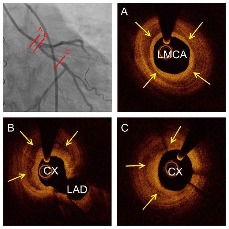
Figure 3.
Intravascular optical coherence tomography at the angiographic lesion sites. (A) Circumflex layered massive organized thrombus in the left main coronary artery (LMCA) covering the ostium of the circumflex (CX) and left anterior descending coronary artery (LAD) (B). Furthermore, crescent-shaped layered organized thrombus was seen in the distal part of the circumflex (C). Yellow arrows show layered thrombosis within the intima vessel layer.

