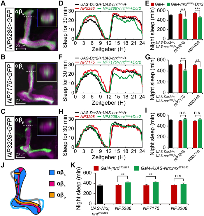
Figure 3. Neurexin functions in αβsc neurons to promote nighttime sleep.
(A–C) Projection views of confocal stacks at the level of the right MB lobes from NP5286-αβs (A), NP7175-αβc (B), and NP3208-αβp (C) flies driving mCD8::GFP (green). In all panels, the αβ lobes are labeled with anti-FasII (magenta). The inset shows a horizontal cross-section through the vertical collateral at the level of the dashed line in each panel. Scale bar = 20 μm. (D,E) Average sleep profiles for UAS-Dicer2/NP5286-GAL4;UAS-nrxRNAi/+ (n = 53) and control (n = 32) flies, plotted as a 30 min moving average (D), and quantification of total nighttime sleep for each genotype (E). Note that Dicer-2 was used to enhance the transgenic RNAi effect in panels D, F, and H. (F,G) Average sleep profiles for NP7175-GAL4/Y;UAS-Dicer2/+;UAS-nrxRNAi/+ (n = 37) and control (n = 32) flies, plotted as a 30 min moving average (F), and quantification of total nighttime sleep for each genotype (G). (H,I) Average sleep profiles for NP3208-GAL4/Y;UAS-Dicer2/+;UAS-nrxRNAi/+ (n = 42) and control (n = 32) flies, plotted as a 30 min moving average (H), and quantification of total nighttime sleep for each genotype (I). (J) Model of the MB αβ neurons illustrating three subsets of neurons within the αβ lobes. Purple, αβc neurons; blue, αβs neurons; yellow, αβp neurons. (K) Total nighttime sleep in flies with rescued neurexin expression using specific GAL4 drivers for each αβ subset. Rescue experiments were conducted in the nrx273/Δ83 background; flies bear one copy of the indicated drivers (n = 16).

